




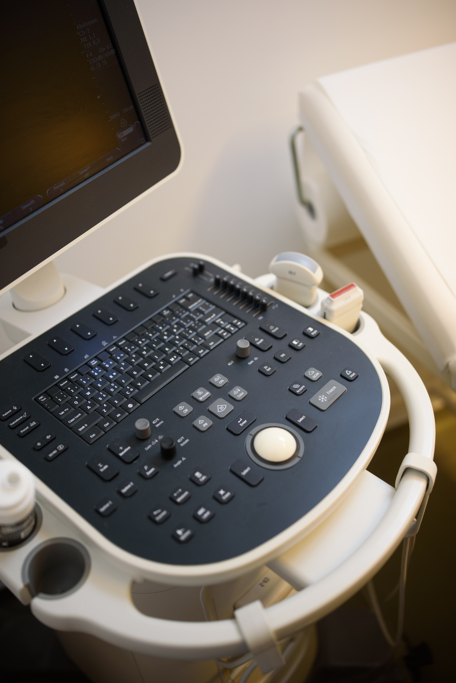
Joint ultrasound
- Dynamic examination We observe how tendons and muscles glide during movement.
- Immediate report You receive the detailed written report at the end of the examination.
- Soft tissue detail Tendons, ligaments, muscles and fluid collections — all become visible.
- Even with a metal prosthesis Where MRI is not an option, ultrasound is a safe alternative.
When is a joint ultrasound recommended?
Joint ultrasound shows the source of pain quickly and without radiation exposure — whether it is inflammation, a tear or a fluid collection.
-
Joint pain and stiffness
For complaints of the shoulder, elbow, wrist, hip, knee or ankle.
-
Swelling and fluid collection
To identify fluid that has built up in the joint capsule.
-
Sports injuries
For torn muscles, strained ligaments and partial sprains.
-
Restricted movement
If a joint does not move through its usual range, or catches.
-
Joint inflammation
To detect changes caused by arthritis and increased blood flow.
-
Tendon sheath inflammation
To show inflammation of the sheath surrounding the tendons.
-
Tunnel syndromes
To check the course of the surrounding nerves and any compression.
-
Rheumatology follow-up
To measure the activity of known conditions and the effectiveness of treatment.
If the pain has lasted for weeks, do not wait any longer: recognising soft tissue injuries early shortens rehabilitation.
This list is for information only and does not replace a specialist examination. Ultrasound shows the condition of the soft tissues — tendons, ligaments, muscles; an X-ray is needed to assess whether the bones are intact. If you have any questions, call us: 06-1-413-2500 (H–P 9–17).
Why choose a joint ultrasound at Endomedix?
-
Dynamic diagnosis during movement
Unlike X-ray or MRI, ultrasound allows the doctor to observe how tendons and muscles glide during movement. This functional approach helps to uncover hidden problems that would remain invisible at rest.
-
Immediate evaluation and report
At the end of the examination, you do not have to wait days for your report. As soon as the measurements are finished, our specialist gives you the detailed written report, explains the changes seen and recommends any further rheumatological or orthopaedic steps.
-
Radiation-free, can be repeated as often as needed
The procedure uses no harmful radiation, so it is completely safe for children, older people and expectant mothers. As it involves no intervention, it is ideal for regular monitoring of the healing process.
How should you prepare for the examination?
Joint ultrasound requires no special diet — there are just a few small things to bear in mind.
-
01
Light clothing
Wear clothing that allows the part of the body being examined — shoulder, knee, ankle — to be uncovered easily.
-
02
Clean skin
Do not use cream or body lotion immediately before the examination, as it reduces image quality.
-
03
Previous reports
Bring your previous X-ray, MRI or ultrasound reports and any discharge summaries relating to the area.
-
04
Cooperation during the examination
The doctor may ask you to make certain movements — these help to get the most accurate images.
Did you know?
Five questions and answers about the examination and the report.
-
Did you know that joint ultrasound also detects hidden inflammation?
In many cases, the cause of pain is not an obvious injury but inflammation of the soft tissues. During the examination, our doctors can show increased blood flow around the joint, which is one of the most reliable signs of active inflammation — this is key to recognising rheumatological conditions early and to setting up targeted treatment.
-
Did you know that ultrasound is one of the most effective tools for detecting a Baker's cyst?
A feeling of tightness behind the knee is often caused by a Baker's cyst, which develops when joint fluid builds up. Ultrasound not only confirms the presence and size of the cyst but also helps to distinguish it from the more dangerous deep vein thrombosis, which can cause similar symptoms.
-
Did you know that ultrasound gives a more accurate picture of a partial ligament tear than an X-ray?
While an X-ray mainly checks whether the bones are intact, it does not show soft tissue changes. Joint ultrasound shows the fibre structure of the ligaments, so it can be determined precisely whether an ankle or knee injury involves a complete or only a partial tear — which fundamentally determines how long rehabilitation will take.
-
Did you know that the examination can also be performed if you have a metal prosthesis or implant?
For many patients, MRI is not an option because of metal in their body, such as a knee or hip prosthesis. Metal does not disturb ultrasound waves to the same extent as magnetic resonance, so this method is an excellent alternative for checking post-operative complications, inflammation around the prosthesis or fluid collections.
-
Did you know that the ultrasound examination can also be paid for from your health fund account?
The fees for diagnostic examinations at Endomedix can be paid in full from your health fund account. This is not only convenient but also financially advantageous: the tax refund on fund contributions can significantly reduce your private healthcare costs.
Can also be paid from a health fund account
The examination fee can of course also be paid from your health fund account — our colleagues will be happy to give you the details. The list of health funds that work with Endomedix can be found here.
- Health fund account The fee for the joint ultrasound and the specialist consultation can also be paid from your health fund account.
- Questions about the details? Call us on +36 1 413 2500 or write to us via the contact page.